Healthcare professionals, neonatal units, hospitals, and health services
Statement of standard
Vascular access is achieved in a competent, skillful and safe manner.
Rationale
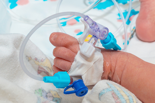
Intravenous (IV) cannulation is among the most common and widespread medical procedures performed on critically ill infants in the NICU. (1) Treatment frequently depends on the use of peripheral or central vascular access devices (VADs) to administer fluids, nutrients, and medication. (2–4) There are several types of VADs, which are inserted into either a vein or an artery. Factors such as body weight, fluid characteristics, availability of venous access sites, and anticipated length of access needed are taken into account when siting a VAD. The frequency of complications, including infiltration/extravasation, leaking, occlusion, thrombosis, and infections, has remained relatively constant over the past 30 years. (5–15)
Benefits
Short-term benefits
Reduced number of skin breaking and painful procedures (16,17)
Reduced occurrence of complications e.g. infections (18)
Long-term benefits
Reduced late consequences of early exposure to antibiotics (consensus)
Reduced risk of long-term consequences of painful procedures for infants and parents (19)
Components of the standard
Component
Grading of evidence
Indicator of meeting the standard
For parents and family
Parents are informed by healthcare professionals about the need and procedure for achieving vascular access.
B (High quality)
Patient information sheet1
Parents are encouraged and guided to comfort the infant if feasible by healthcare professionals. (20) (see Care procedures)
B (High quality)
Patient information sheet1
For healthcare professionals
A unit guideline on the aseptic insertion and maintenance of vascular access devices (VADs) is adhered to by all healthcare professionals. (21)
A (High quality) B (High quality)
Guideline
The necessity for ongoing vascular access is identified.
B (High quality)
Guideline
The procedure is approached in a developmentally supportive manner using (none)-pharmacological pain relieving treatment. (10,22–26) (see Infant- and family-centred developmental care)
A (Moderate quality) B (Moderate quality)
Guideline
Training on the insertion of VADs is attended by all responsible healthcare professionals.
B (High quality)
Training documentation
For neonatal unit
A unit guideline on the aseptic insertion and maintenance of VADs is available and regularly updated.
B (High quality)
Guideline
For hospital
Training on the aseptic insertion of VADs is ensured.
B (High quality)
Training documentation
Equipment to administer and monitor infusion therapy is suitable for a neonatal population.
B (High quality)
Audit report2
For health service
A national guideline on the aseptic insertion and maintenance of VADs is available and regularly updated.
B (High quality)
Guideline
1The indicator “patient information sheet” is an example for written, detailed information, in which digital solutions are included, such as web-based systems, apps, brochures, information leaflets, and booklets.
2The indicator “audit report” can also be defined as a benchmarking report.
Where to go
Further development
Grading of evidence
For parents and family
N/A
For healthcare professionals
N/A
For neonatal unit and hospital
Optimise the use of specially trained vascular access professionals.
A (Low quality) B (Moderate quality)
For health service
Develop a European Vascular Access Certification programme for all healthcare professionals in the field.
B (Moderate quality)
Getting started
Initial steps
For parents and family
Parents are verbally informed by healthcare professionals about the need and procedure for achieving vascular access.
If present, parents are invited to support their infant before, during and after the insertion of vascular access devices (VADs).
For healthcare professionals
Attend training on the aseptic insertion and maintenance of VADs.
For neonatal unit
Develop and implement a unit guideline on the aseptic insertion and maintenance of VADs.
Provide a flow chart that guarantees most appropriate Vascular Access Device to meet each infant’s current and anticipated needs. (23)
Provide a vascular visualisation devise for vascular assessment and insertion support if required.
Conduct data collection and compliance monitoring.
Develop information material for parents on the need and procedure for achieving vascular access. (10,24,25)
For hospital
Support healthcare professionals to participate in training on peripheral and central venous/arterial access.
Provide a vascular visualisation device for vascular assessment and insertion support if required.
For health service
Develop and implement a national guideline on the aseptic insertion and maintenance of VADs including indication for insertion, type of device, access visualisation, and management of access and complications.
Zempsky WT. Optimizing the management of peripheral venous access pain in children: evidence, impact, and implementation. Pediatrics. 2008 Nov;122 Suppl 3:S121-124.
Millam DA. Managing complications of i.v. therapy (continuing education credit). Nursing (Lond). 1988 Mar;18(3):34–43.
Carbajal R, Rousset A, Danan C, Coquery S, Nolent P, Ducrocq S, et al. Epidemiology and treatment of painful procedures in neonates in intensive care units. JAMA. 2008 Jul 2;300(1):60–70.
Pettit J. Assessment of the infant with a peripheral intravenous device. Adv Neonatal Care Off J Natl Assoc Neonatal Nurses. 2003 Oct;3(5):230–40.
Franck LS, Hummel D, Connell K, Quinn D, Montgomery J. The safety and efficacy of peripheral intravenous catheters in ill neonates. Neonatal Netw NN. 2001 Aug;20(5):33–8.
Batton DG, Maisels MJ, Appelbaum P. Use of peripheral intravenous cannulas in premature infants: a controlled study. Pediatrics. 1982 Sep;70(3):487–90.
Reynolds J. Comparison of percutaneous venous catheters and teflon catheters for intravenous therapy in neonates. Neonatal Netw NN. 1993 Aug;12(5):33–9.
Stanley MD, Meister E, Fuschuber K. Infiltration during intravenous therapy in neonates: comparison of Teflon and Vialon catheters. South Med J. 1992 Sep;85(9):883–6.
Sheehan AM, Palange K, Rasor JS, Moran MA. Significantly improved peripheral intravenous catheter performance in neonates: insertion ease, dwell time, complication rate, and costs. J Perinatol Off J Calif Perinat Assoc. 1992 Dec;12(4):369–76.
Johnston C, Campbell-Yeo M, Disher T, Benoit B, Fernandes A, Streiner D, et al. Skin-to-skin care for procedural pain in neonates. Cochrane Neonatal Group, editor. Cochrane Database Syst Rev [Internet]. 2017 Feb 16 [cited 2018 May 8]; Available from: http://doi.wiley.com/10.1002/14651858.CD008435.pub3
Tobin CR. The Teflon intravenous catheter: incidence of phlebitis and duration of catheter life in the neonatal patient. J Obstet Gynecol Neonatal Nurs JOGNN. 1988 Feb;17(1):35–42.
Collinge JM, Aranda JV. Nonmetabolic complications of neonatal intravenous therapy: epidemiologic considerations. Am J Perinatol. 1984 Jan;1(2):185–9.
Phelps SJ, Cochran EB. Effect of the continuous administration of fat emulsion on the infiltration of intravenous lines in infants receiving peripheral parenteral nutrition solutions. JPEN J Parenter Enteral Nutr. 1989 Dec;13(6):628–32.
Hecker JF. Failure of intravenous infusions from extravasation and phlebitis. Anaesth Intensive Care. 1989 Nov;17(4):433–9.
Webb AA. Methods of intravenous therapy in preterm infants. Issues Compr Pediatr Nurs. 1987;10(4):215–21.
Ainsworth S, McGuire W. Percutaneous central venous catheters versus peripheral cannulae for delivery of parenteral nutrition in neonates. Cochrane Database Syst Rev. 2015 Oct 6;(10):CD004219.
Ainsworth SB, McGuire W. Peripherally Inserted Central Catheters vs Peripheral Cannulas for Delivering Parenteral Nutrition in Neonates. JAMA. 2016 Jun 21;315(23):2612–3.
Barría RM, Lorca P, Muñoz S. Randomized controlled trial of vascular access in newborns in the neonatal intensive care unit. J Obstet Gynecol Neonatal Nurs JOGNN. 2007 Oct;36(5):450–6.
Grunau RE. Neonatal pain in very preterm infants: long-term effects on brain, neurodevelopment and pain reactivity. Rambam Maimonides Med J. 2013;4(4):e0025.
Phipps K, Modic A, O’Riordan MA, Walsh M. A randomized trial of the Vein Viewer versus standard technique for placement of peripherally inserted central catheters (PICCs) in neonates. J Perinatol Off J Calif Perinat Assoc. 2012 Jul;32(7):498–501.
Hartley KA, Miller CS, Gephart SM. Facilitated tucking to reduce pain in neonates: evidence for best practice. Adv Neonatal Care Off J Natl Assoc Neonatal Nurses. 2015 Jun;15(3):201–8.
Catelin C, Tordjman S, Morin V, Oger E, Sizun J. Clinical, physiologic, and biologic impact of environmental and behavioral interventions in neonates during a routine nursing procedure. J Pain Off J Am Pain Soc. 2005 Dec;6(12):791–7.
Vinall J, Grunau RE. Impact of repeated procedural pain-related stress in infants born very preterm. Pediatr Res. 2014 May;75(5):584–7.
Vinall J, Miller SP, Bjornson BH, Fitzpatrick KPV, Poskitt KJ, Brant R, et al. Invasive Procedures in Preterm Children: Brain and Cognitive Development at School Age. PEDIATRICS. 2014 Mar 1;133(3):412–21.
Stevens B, Yamada J, Ohlsson A, Haliburton S, Shorkey A. Sucrose for analgesia in newborn infants undergoing painful procedures. Cochrane Neonatal Group, editor. Cochrane Database Syst Rev [Internet]. 2016 Jul 15 [cited 2018 May 8]; Available from: http://doi.wiley.com/10.1002/14651858.CD001069.pub5
November 2018 / 1st edition / next revision: 2023
Recommended citation
EFCNI, Van Rens R, Helder, O et al., European Standards of Care for Newborn Health: Vascular access. 2018.
Please enter your job description and origin country
You are currently viewing a placeholder content from Facebook. To access the actual content, click the button below. Please note that doing so will share data with third-party providers.
You are currently viewing a placeholder content from Instagram. To access the actual content, click the button below. Please note that doing so will share data with third-party providers.
You are currently viewing a placeholder content from X. To access the actual content, click the button below. Please note that doing so will share data with third-party providers.