Term and preterm infants at risk for brain injury:Infants with hypoxic-ischaemic encephalopathy (HIE)
Infants with encephalopathy for other causes (e.g. metabolic)
Infants with suspected or verified seizures
Infants requiring intensive care and/or surgery
Infants with suspected/confirmed congenital central nervous system (CNS) anomalies
Parents
User group
Healthcare professionals, neonatal units, hospitals, and health services
Statement of standard
In order to improve evaluation and outcomes of newborn infants at risk of brain injury, management includes neurological monitoring using a structured, age-appropriate neurological assessment and a range of devices to evaluate brain haemodynamics, oxygen transport, brain function, and imaging, as required.
Rationale
Newborn infants comprise a high-risk population for developing brain injury, during the first days after birth due to respiratory, haemodynamic, infectious, or metabolic instability. Full term and preterm infants with hypoxia-ischaemia, CNS infections, or congenital anomalies are at particular risk of brain injury. Early recognition of on-going disturbances of brain function or structural damage is important in implementing preventive or treatment strategies, and appropriate follow-up. Early detection of cerebral compromise, such as encephalopathy and seizures, is associated with better management of these conditions. High-risk infants should be identified as early as possible, the patient history together with a structured clinical examination and repeated clinical observations form the basis of the evaluation. The electroencephalogram (EEG) provides sensitive detection of abnormal brain function. (1,2) Continuous monitoring with a full montage EEG or the limited-channel amplitude-integrated EEG (aEEG) has been increasingly used in neonatal units and is excellent in the detection and grading of the severity of cerebral compromise in both term and preterm infants, and can be used for early evaluation before interventions such as therapeutic hypothermia. (3–11) Modern aEEG monitors also display the raw EEG (aEEG/EEG), which improves seizure detection. (12,13) The use of continuous EEG or aEEG/EEG monitoring is associated with earlier seizure diagnosis and better seizure management. (14,15) Studies in asphyxiated newborn infants have shown that aEEG combined with near-infrared spectroscopy is useful. (16,17)
Benefits
Short-term benefits
Improved evaluation of clinical symptoms, including seizures, and early detection of cerebral compromise (2,6,14,18,19)
Refined clinical management of neonatal seizures, including more efficient treatment and less use of antiepileptic drugs (14,15,18–20)
Early prediction of outcome may assist medical decisions such as interventions and redirection of care (5,9,10,21)
Long-term benefits
Improved long-term outcomes (22–25)
Improved cost-effectiveness (26,27)
Reduced exposure to antiepileptic drugs (15,20)
Components of the standard
Component
Grading of evidence
Indicator of meeting the standard
For parents and family
Parents are informed by healthcare professionals about the role of EEG and aEEG/EEG monitoring.
B (High quality)
Patient information sheet1
For healthcare professionals
A unit guideline on neurological monitoring including EEG and aEEG/EEG monitoring is adhered to by all healthcare professionals, to include
asphyxiated newborn infants, including undergoing therapeutic hypothermina (3–5,5,6,16,23,26)
infants at risk of and undergoing treatment for seizures. (6,12–15,18–20,22,23,28)
A (High quality) B (High quality)
Audit report2, guideline
Specific training on the use of EEG and aEEG/EEG monitoring is attended by all responsible healthcare professionals. (5,11,14,27,29)
A (Moderate quality) B (High quality)
Training documentation
Teams with a focus of interest on EEG and aEEG/EEG monitoring (e.g. neonatologists, neurologists, neuro-physiologists, nurses, radiologists, radiographers, and physicists) are established. (29,30)
A (High quality) B (High quality)
Guideline
For neonatal unit
A unit guideline on the implications of EEG and aEEG/EEG monitoring is available and regularly updated.
B (High quality)
Guideline
For hospital
Training on the use of EEG and aEEG/EEG monitoring is ensured. (5,11,14,27,29)
A (Moderate quality) B (High quality)
Guideline
An interdisciplinary team for neurological evaluation (including EEG and aEEG/EEG) of high-risk infants in the NICU is supported. (14,15)
A (Moderate quality)
Audit report2
Facilities for EEG and aEEG/EEG monitoring and interpretation are provided.
B (High quality)
Audit report2
For health service
High-risk infants are transferred to NICUs with appropriate neuro-monitoring systems and expertise. (31)
A (High quality)
Audit report2, guideline
1The indicator “patient information sheet” is an example for written, detailed information, in which digital solutions are included, such as web-based systems, apps, brochures, information leaflets, and booklets.
2The indicator “audit report” can also be defined as a benchmarking report.
Where to go
Further development
Grading of evidence
For parents and family
N/A
For healthcare professionals
N/A
For neonatal unit
Develop a full neonatal neuro-critical care concept, including guidelines and close collaboration with neurologists. (13,19,24)
A (Moderate quality)
For hospital
Use monitoring systems that allow for expert evaluation of aEEG/EEG or EEG 24/7 also from outside the hospital.
A (Low quality)
For health service
Monitor incidence, treatment and long-term outcomes after neonatal seizures. (15,24,25)
A (High quality)
Develop multi-centre expertise by sharing EEG databases.
B (Moderate quality)
Getting started
Initial steps
For parents and family
Parents are verbally informed by healthcare professionals about the implications of EEG monitoring.
For healthcare professionals
Attend training on the use of EEG and aEEG/EEG monitoring.
Identify leading staff with a focus of interest on neonatal neurological evaluation and monitoring.
For neonatal unit
Develop and implement a unit guideline on the use of EEG and aEEG/EEG monitoring.
Develop parental information material about EEG and aEEG/EEG monitoring, also including parental perspectives.
Provide resources for specific training on EEG and aEEG/EEG monitoring tools.
For hospital
Support healthcare professionals to participate in training on the use of aEEG/EEG and EEG monitoring.
Provide technology for EEG or aEEG/EEG monitoring.
For health service
Create systems to effectively transfer high-risk infants to NICUs with appropriate neuromonitoring systems and expertise.
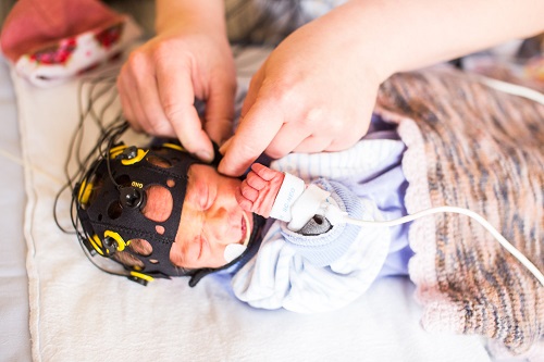
Electroencephalography (EEG) and amplitude-integrated EEG (aEEG) for evaluation of brain function in high risk infants
A majority of adverse events affecting brain function in term and preterm infants occur during deliver or the first week after birth. Such events include perinatal asphyxia, cerebral haemorrhages, ischaemia, metabolic and infectious conditions. Cerebral symptoms may be vague or entirely absent but may also include encephalopathy and seizures. Evaluation of brain function with convetional EEG or continuous monitoring with the aEEG/EEG gives diagnostic and prognostic information in high-risk term and preterm infants. Continuous video-EEG monitoring can be considered to be the gold standard, but this method is not available in all neonatal intensive care units (NICU) and not feasible for routine monitoring of large numbers of high-risk infants. During the last two decades EEG and aEEG/EEG monitoring have been increasingly used worldwide in compromised newborn infants.
Newborn infants with compromised brain function or at risk for developing severe cerebral complications should be monitored closely by clinical observation and continuously with aEEG/EEG. Conventional EEG should be performed in newborn infants monitored by aEEG/EEG.
Regular knowledge updates and training of healthcare professionals in basic management and evaluation of aEEG/EEG and EEG is of utmost importance. Several studies have reported that insufficient training in aEEG/EEG is associated with poorer and unreliable performance of the monitoring. For this reason, aEEG/EEG monitoring should be conducted in collaboration with clinical neurophysiologists or neurologists, and for most monitored newborn infants at least one standard EEG should be recorded. (13,28–30,32–35)
Several studies have demonstrated that the electrocortical activity is one of the most sensitive measures for early evaluation of brain function and early prediction of outcome in asphyxiated newborn infants. Consequently, it is recommended to record the aEEG/EEG for evaluation of asphyxiated newborn infants before hypothermia treatment. (5,21) In asphyxiated term infants, simultaneous monitoring with aEEG/EEG and NIRS is associated with more precise outcome prediction. (16,17)
Clinical identification of suspected seizures is not reliable since a majority of neonatal seizures have only subtle clinical symptoms or are entirely subclinical. Brain monitoring with aEEG/EEG and EEG in asphyxiated newborn infants allows earlier recognition of seizures in newborn infants with HIE, and with more precise treatment of seizures and the use of fewer antiepileptic drugs. (14,15,18,19,22–25,36)
The compressed aEEG trend alone is not sensitive enough for detection of seizures since especially brief seizures may be missed in the compressed trend. However, if both aEEG and raw-EEG is inspected around 80-90% of all seizures that can be identified in a standard EEG may be detected. (13,37) Development of efficient automated seizure detection alarms is urgently needed in the busy NICU setting.
Many studies have also shown that aEEG and EEG may be sensitive early predictors of outcome in preterm infants (1,7,9,10), but clinical experience of aEEG in very preterm infants is still limited. The early predictive accuracy of aEEG and EEG can be expected to be lower in preterm infants than in term infants since the long-term outcome of especially very preterm infants may be affected by later complications during the clinical course.
The neonatal neurocritical care concept is an emerging strategy which includes a care concept based on specially trained NICU healthcare professionals and interdisciplinary teams that include neurologists, guidelines and protocols for consistent management of newborn infants at risk of neurological injury, aEEG/EEG and EEG monitoring of high-risk infants, and long-term follow up. (31) The first reports from NICUs practicing neurocritical care show very promising results. (15,24,25,27)
Watanabe K, Hayakawa F, Okumura A. Neonatal EEG: a powerful tool in the assessment of brain damage in preterm infants. Brain Dev. 1999 Sep;21(6):361–72.
Lamblin MD, Walls EE, André M. The electroencephalogram of the full-term newborn: review of normal features and hypoxic-ischemic encephalopathy patterns. Neurophysiol Clin Clin Neurophysiol. 2013 Dec;43(5–6):267–87.
Hellström-Westas L, Rosén I, Svenningsen NW. Predictive value of early continuous amplitude integrated EEG recordings on outcome after severe birth asphyxia in full term infants. Arch Dis Child Fetal Neonatal Ed. 1995 Jan;72(1):F34-38.
Shalak LF, Laptook AR, Velaphi SC, Perlman JM. Amplitude-integrated electroencephalography coupled with an early neurologic examination enhances prediction of term infants at risk for persistent encephalopathy. Pediatrics. 2003 Feb;111(2):351–7.
Thoresen M, Hellström-Westas L, Liu X, de Vries LS. Effect of hypothermia on amplitude-integrated electroencephalogram in infants with asphyxia. Pediatrics. 2010 Jul;126(1):e131-139.
Nash KB, Bonifacio SL, Glass HC, Sullivan JE, Barkovich AJ, Ferriero DM, et al. Video-EEG monitoring in newborns with hypoxic-ischemic encephalopathy treated with hypothermia. Neurology. 2011 Feb 8;76(6):556–62.
Hellström-Westas L, Klette H, Thorngren-Jerneck K, Rosén I. Early prediction of outcome with aEEG in preterm infants with large intraventricular hemorrhages. Neuropediatrics. 2001 Dec;32(6):319–24.
Olischar M, Klebermass K, Waldhoer T, Pollak A, Weninger M. Background patterns and sleep-wake cycles on amplitude-integrated electroencephalography in preterms younger than 30 weeks gestational age with peri-/intraventricular haemorrhage. Acta Paediatr Oslo Nor 1992. 2007 Dec;96(12):1743–50.
Fogtmann EP, Plomgaard AM, Greisen G, Gluud C. Prognostic Accuracy of Electroencephalograms in Preterm Infants: A Systematic Review. Pediatrics. 2017 Feb;139(2).
Jiang C-M, Yang Y-H, Chen L-Q, Shuai X-H, Lu H, Xiang J-H, et al. Early amplitude-integrated EEG monitoring 6 h after birth predicts long-term neurodevelopment of asphyxiated late preterm infants. Eur J Pediatr. 2015 Aug;174(8):1043–52.
Murray DM, Boylan GB, Ryan CA, Connolly S. Early EEG Findings in Hypoxic-Ischemic Encephalopathy Predict Outcomes at 2 Years. Pediatrics. 2009 Sep 1;124(3):e459–67.
Shellhaas RA, Soaita AI, Clancy RR. Sensitivity of amplitude-integrated electroencephalography for neonatal seizure detection. Pediatrics. 2007 Oct;120(4):770–7.
Zhang L, Zhou Y-X, Chang L-W, Luo X-P. Diagnostic value of amplitude-integrated electroencephalogram in neonatal seizures. Neurosci Bull. 2011 Aug;27(4):251–7.
Shellhaas R, Barks A. Impact of amplitude-integrated EEG on the clinical care for neonates with seizures. Pediatr Neurol. 2012 Jan;46(1):32–5.
Wietstock SO, Bonifacio SL, McCulloch CE, Kuzniewicz MW, Glass HC. Neonatal Neurocritical Care Service Is Associated With Decreased Administration of Seizure Medication. J Child Neurol. 2015 Aug;30(9):1135–41.
Toet MC, Lemmers PMA, Schelven LJ van, Bel F van. Cerebral Oxygenation and Electrical Activity After Birth Asphyxia: Their Relation to Outcome. Pediatrics. 2006 Feb 1;117(2):333–9.
Lemmers PMA, Zwanenburg RJ, Benders MJNL, de Vries LS, Groenendaal F, van Bel F, et al. Cerebral oxygenation and brain activity after perinatal asphyxia: does hypothermia change their prognostic value? Pediatr Res. 2013 Aug;74(2):180–5.
Murray DM, Boylan GB, Ali I, Ryan CA, Murphy BP, Connolly S. Defining the gap between electrographic seizure burden, clinical expression and staff recognition of neonatal seizures. Arch Dis Child Fetal Neonatal Ed. 2008 May;93(3):F187-191.
Glass HC, Shellhaas RA, Wusthoff CJ, Chang T, Abend NS, Chu CJ, et al. Contemporary Profile of Seizures in Neonates: A Prospective Cohort Study. J Pediatr. 2016 Jul;174:98–103.e1.
Hellström-Westas L, Blennow G, Lindroth M, Rosén I, Svenningsen NW. Low risk of seizure recurrence after early withdrawal of antiepileptic treatment in the neonatal period. Arch Dis Child Fetal Neonatal Ed. 1995 Mar;72(2):F97-101.
Bonifacio SL, deVries LS, Groenendaal F. Impact of hypothermia on predictors of poor outcome: how do we decide to redirect care? Semin Fetal Neonatal Med. 2015 Apr;20(2):122–7.
van Rooij LGM, Toet MC, van Huffelen AC, Groenendaal F, Laan W, Zecic A, et al. Effect of treatment of subclinical neonatal seizures detected with aEEG: randomized, controlled trial. Pediatrics. 2010 Feb;125(2):e358-366.
Kharoshankaya L, Stevenson NJ, Livingstone V, Murray DM, Murphy BP, Ahearne CE, et al. Seizure burden and neurodevelopmental outcome in neonates with hypoxic-ischemic encephalopathy. Dev Med Child Neurol. 2016 Dec;58(12):1242–8.
Bashir RA, Espinoza L, Vayalthrikkovil S, Buchhalter J, Irvine L, Bello-Espinosa L, et al. Implementation of a Neurocritical Care Program: Improved Seizure Detection and Decreased Antiseizure Medication at Discharge in Neonates With Hypoxic-Ischemic Encephalopathy. Pediatr Neurol. 2016 Nov;64:38–43.
Harris ML, Malloy KM, Lawson SN, Rose RS, Buss WF, Mietzsch U. Standardized Treatment of Neonatal Status Epilepticus Improves Outcome. J Child Neurol. 2016;31(14):1546–54.
Gray J, Geva A, Zheng Z, Zupancic JAF. CoolSim: using industrial modeling techniques to examine the impact of selective head cooling in a model of perinatal regionalization. Pediatrics. 2008 Jan;121(1):28–36.
Massaro AN, Murthy K, Zaniletti I, Cook N, DiGeronimo R, Dizon MLV, et al. Intercenter Cost Variation for Perinatal Hypoxic-Ischemic Encephalopathy in the Era of Therapeutic Hypothermia. J Pediatr. 2016 Jun;173:76–83.e1.
Zhang D, Liu Y, Hou X, Zhou C, Luo Y, Ye D, et al. Reference Values for Amplitude-Integrated EEGs in Infants From Preterm to 3.5 Months of Age. Pediatrics. 2011 May 1;127(5):e1280–7.
Griesmaier E, Neubauer V, Ralser E, Trawöger R, Kiechl-Kohlendorfer U, Keller M. Need for quality control for aEEG monitoring of the preterm infant: a 2-year experience. Acta Paediatr Oslo Nor 1992. 2011 Aug;100(8):1079–83.
Sacco L. Amplitude-Integrated Electroencephalography Interpretation During Therapeutic Hypothermia: An Educational Program and Novel Teaching Tool. Neonatal Netw NN. 2016;35(2):78–86.
Glass HC, Bonifacio SL, Peloquin S, Shimotake T, Sehring S, Sun Y, et al. Neurocritical care for neonates. Neurocrit Care. 2010 Jun;12(3):421–9.
Hellström-Westas L, Rosén I, Vries L, Greisen G. Amplitude-integrated EEG Classification and Interpretation in Preterm and Term Infants. NeoReviews. 2006 Feb 1;7.
André M, Lamblin M-D, d’Allest AM, Curzi-Dascalova L, Moussalli-Salefranque F, S Nguyen The T, et al. Electroencephalography in premature and full-term infants. Developmental features and glossary. Neurophysiol Clin Clin Neurophysiol. 2010 May;40(2):59–124.
Lamblin MD, de Villepin-Touzery A. EEG in the neonatal unit. Neurophysiol Clin Clin Neurophysiol. 2015 Mar;45(1):87–95.
Shellhaas RA, Chang T, Tsuchida T, Scher MS, Riviello JJ, Abend NS, et al. The American Clinical Neurophysiology Society’s Guideline on Continuous Electroencephalography Monitoring in Neonates. J Clin Neurophysiol Off Publ Am Electroencephalogr Soc. 2011 Dec;28(6):611–7.
Boylan G, Rennie J, Pressler R, Wilson G, Morton M, Binnie C. Phenobarbitone, neonatal seizures, and video-EEG. Arch Dis Child Fetal Neonatal Ed. 2002 May;86(3):F165–70.
Shellhaas RA, Clancy RR. Characterization of neonatal seizures by conventional EEG and single-channel EEG. Clin Neurophysiol. 2007 Oct 1;118(10):2156–61.
November 2018 / 1st edition / next revision: 2023
Recommended citation
EFCNI, Hellström-Westas L, Zimmermann LJI et al., European Standards of Care for Newborn Health: Neurologic monitoring of the high-risk infant: EEG and aEEG. 2018
Please enter your job description and origin country
You are currently viewing a placeholder content from Facebook. To access the actual content, click the button below. Please note that doing so will share data with third-party providers.
You are currently viewing a placeholder content from Instagram. To access the actual content, click the button below. Please note that doing so will share data with third-party providers.
You are currently viewing a placeholder content from X. To access the actual content, click the button below. Please note that doing so will share data with third-party providers.