Term and preterm infants at risk for brain injury:Infants with hypoxic-ischaemic encephalopathy (HIE)
Infants with encephalopathy for other causes (e.g. metabolic)
Infants with suspected or verified seizures
Infants requiring intensive care and/or surgery
Infants with suspected/confirmed congenital central nervous system (CNS) anomalies
Parents
User group
Healthcare professionals, neonatal units, hospitals, and health services
Statement of standard
In order to improve evaluation and outcomes of newborn infants at risk of brain injury, management includes neurological monitoring using a structured, age-appropriate neurological assessment and a range of devices to evaluate brain haemodynamics, oxygen transport, brain function, and imaging, as required.
Rationale
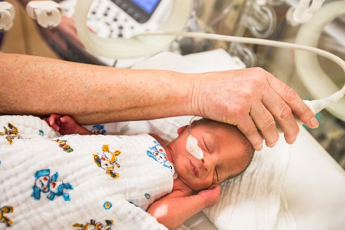
Infants requiring neonatal intensive care constitute a high-risk population for developing brain injury, especially during the first days after birth due to respiratory, haemodynamic, infectious, or metabolic instability. Full term and preterm infants exposed to hypoxia-ischaemia or infections, or carrying conditions such as congenital malformations, antenatal (maternal) risk factors, neonatal diseases potentially involving CNS, or late prematurity, among others, are exposed to increased risk of brain injury. Early recognition of on-going disturbances of brain function or structural damage is important in implementing preventive or treatment strategies, and appropriate follow-up. Early detection of cerebral compromise, such as encephalopathy or seizures, is associated with better management of these conditions. High-risk infants should be identified as early as possible, the patient history together with a structured clinical examination and repeated clinical observations form the basis of the evaluation. The vulnerability of this population, the severity of underlying clinical conditions, and the complexity of care deserve preferably continuous, cot-side, and non-invasive monitoring tools. This can be accomplished from four perspectives: haemodynamics and oxygen transport, connectivity and function, structure, and clinical expression. The ultimate goal is to prevent or reduce risk for brain injury by early identification of high-risk infants and improved clinical management.
Benefits
Short-term benefits
Reduced mortality and morbidity (i.e., detect sinovenous thrombosis, severe haemorraghes or post-haemorrhagic ventricular dilatation) (1–11)
Direct feedback on neuroprotective interventions (i.e., low molecular weight heparin treatment for cerebral vein thrombosis, ventricular reservoir taps, ventriculo-peritoneal shunt treatment) (1–8)
Improved assessment of severity of brain damage which might redirect care (i.e., in patients with hypoxic ischaemic encephalopathy (HIE), arterial stroke, venous infarction) (1–12)
Provides proxy biomarker for outcome for evaluation in neuroprotective intervention trials (11–16)
Informs prognosis for physicians and parents (11–16)
Long-term benefits
More focused follow-up programmes (5,16–20)
Improved understanding of brain injury pathophysiology (5,12,14,17–19)
Improved assessment of neonatal brain development to guide future prevention and intervention strategies (5,16–19)
Components of the standard
Component
Grading of evidence
Indicator of meeting the standard
For parents and family
Parents are informed by healthcare professionals about the role of brain imaging. (21)
A (Moderate quality) B (High quality)
Patient information sheet1
For healthcare professionals
A unit guideline on neurological monitoring including brain imaging is adhered to by all healthcare professionals, to include
term infants with suspected brain injury (1–5,9–13,15,16)
very preterm infants (1–5,12)
A (High quality) B (High quality)
Audit report2, guideline
Training on ultrasound and MRI procedures is attended by all responsible healthcare professionals.
B (High quality)
Training documentation
Teams with a focus of interest on neuroimaging (e.g. nurses, neonatologists, neurologists, neuro-physiologists, radiologists, radiographers, and physicists) are established. (18)
B (High quality)
Guideline
For neonatal unit
A unit guideline on neurological monitoring including brain imaging is available and regularly updated, to include standardised operational procedures for cranial ultrasound (CUS) (22–24) and magnetic resonance imaging (MRI). (20,21,25–27)
A (High quality) B (High quality)
Guideline
For hospital
Training on ultrasound and MRI procedures is ensured. (20,21,25–27)
A (High quality) B (High quality)
Training documentation
An interdisciplinary team for neurological evaluation of high-risk infants in the NICU is supported.
B (Moderate quality)
Audit report2
Facilities for brain imaging (CUS and MRI) are provided.
B (High quality)
Audit report2
For health service
High-risk infants are transferred to NICUs with appropriate neuro-monitoring systems and expertise. (17,28)
A (High quality)
Audit report2, guideline
1The indicator “patient information sheet” is an example for written, detailed information, in which digital solutions are included, such as web-based systems, apps, brochures, information leaflets, and booklets.
2The indicator “audit report” can also be defined as a benchmarking report.
Where to go
Further development
Grading of evidence
For parents and family
N/A
For healthcare professionals
N/A
For neonatal unit
Develop a full neonatal neuro-critical care concept, including guidelines and close collaboration with neurologists.
For hospital
N/A
For health service
Monitor incidence, treatment and long-term outcomes after neonatal brain injury such as intra-ventricular haemorrhage. (18)
A (High quality)
Develop multi-centre expertise by sharing imaging databases.
B (Moderate quality)
Getting started
Initial steps
For parents and family
Parents are verbally informed by healthcare professionals about the role of brain imaging.
For healthcare professionals
Attend training on ultrasound and magnetic resonance imaging (MRI) procedures.
Identify leading healthcare professionals with a focus of interest on neonatal neurological monitoring.
For neonatal unit
Develop and implement a unit guideline on neurological monitoring including brain imaging.
Develop parental information material about brain imaging, also including parental perspectives.
Provide resources for specific training on brain imaging tools.
For hospital
Support healthcare professionals to participate in training on ultrasound and MRI procedures.
For health service
Create systems to effectively transfer high-risk infants to NICUs with appropriate neuro-monitoring systems and expertise.
Despite several major advances in fetal and neonatal care, the frequency of neurodevelopmental disability among the survivors of neonatal intensive care remains high. Although mortality for both, preterm infants and severely compromised term infants has decreased, the population of newborn infants at risk for neurological disability is still increasing. (29,30) Neuroimaging is a critical investigation in the provision of adequate diagnostic or prognostic information for parents. (1–5) Neuroimaging in newborn infants at risk of brain damage is oriented to: a. Diagnosing brain injury to provide the most appropriate medical management. b. Early detection of lesions associated with long-term neurodevelopmental disabilities.
Early diagnosis of structural brain damage can steer neuroprotective and/or neurorehabilitation treatment strategies, and guide appropriate follow up. It can also give us an understanding of the pathophysiology. (1–5)
Neonatal neuroimaging techniques such as CUS, MRI and CT scanning have been used for many decades and have proven to be extremely helpful assessing brain maturation and injury. However, there are still several challenges associated with neonatal neuroimaging, which will be highlighted below. (5,11,13,25)
Proper assessment of neonatal brain images requires extensive knowledge about neonatal brain injury (aetiology, pathophysiology, prognosis), developmental neuro-anatomy (neuro-embryology), the advantages and disadvantages of the different imaging techniques, pitfalls and optimal timing. (5,11,13,25) Furthermore, the transport and sedation of critically ill neonates for both MRI and CT scanning often represents a major challenge. (25–27) Proper scanning requires a dedicated team. The most common used neonatal neuroimaging modalities are: CUS and MRI. The use of CT is very limited and because of radiation should tried to be avoided. All these factors have to be taken into account when choosing timing and modality to image the neonatal brain.
There are advantages and disadvantages for each of the modalities: (5,11,13,25–27)
Cerebral Ultrasound
Advantages
Bedside, patient friendly, save
Reliable for detection of severe haemorrhagic lesions (e.g. peri- intraventricular haemorrhage-P/IVH- in preterms) and severe white matter damage
Doppler technique (detection of thrombosis)
Specific lesions: germinolytic cysts, calcifications, lenticulo-strtriate vasculopathy -LSV
Repeated assessments (i.e., measurements such as the Levene index in posthaemorrhagic ventricle dilatation –PHVD patients)
Disadvantages
Difficult to detect cortical abnormalities
Difficult to detect posterior fossa abnormalities (the use of posterior fontanel and mastoid fontanel can be of help)
Less reliable in detecting small lesions and subtle white matter damage
Difficult to assess myelinisation
MRI
Advantages
Relatively safe (no radiation)
Good assessment of whole brain (including cortex and posterior fossa)
Detailed information about brain development (including myelination)
Reliable information about localisation and extent of brain damage
Reliable for detection of small lesions and subtle white matter damage
Special sequences for different purposes: e.g.,diffusion-weighted imaging (DWI) (cytotoxic oedema), diffusion tensor imaging (DTI) (quantitative white matter tract analysis), magnetic resonance venography (MRV) (venous system), susceptibility weighted imaging (SWI) (haemorrhages), contrast (tumour, abscess), magnetic resonance angiography (MRA) (arterial vessels)
Disadvantages
More burden/distress for (often unstable) infants and medical team: transport issues, sedation, time consuming
High costs (depending on hospital)
Some lesions more difficult to assess (LSV, calcifications, germinolytic cysts)
CT
Advantages
Good visualisation of bone structures
Often wider availability than MRI
Disadvantages
Relatively unsafe (radiation)
Poor tissue contrast (low resolution)
To detect haemorrhages beyond one week can be difficult
Disease states and recommended neuroimaging technique*:
*Based on several factors, including local availability, expertise, and protocols.
Cerebral Ultrasound (examples)
High-risk neonatal conditions: e.g. preterms (gestational age (GA) less than 32 weeks), intrauterine growth restriction, congenital abnormalities (syndromes), post-resuscitation, HIE, meningitis/encephalitis, metabolic diseases, symptomatic hypoglycaemia, hyperbilirubinemia (above exchange transfusion threshold), sudden severe anaemia, congenital heart defects, post-surgery, pre-extracorporeal membrane oxygenation (ECMO), post-ECMO, sudden clinical deterioration.
Newborn infants with neurological symptoms/signs: e.g. seizures, hyper- or hypotonia, abnormal movements, abnormal consciousness, unexplained central apneas, unexplained irritability and restlessness, micro- or macrocephaly.
MRI (examples)
Neurological symptoms not explained by other diagnoses
Convulsions
Symptomatic hypoglycaemia
Severe hyperbilirubinemia and neurological symptoms or abnormal ultrasound
HIE grade II or III
P/IVH with PHVD or periventricular haemorrhagic infarction (PVHI)
Neurological symptoms suggesting brain injury: as soon as possible (to exclude acute conditions that need intervention)
Suspected congenital CNS abnormalities: 1st day after birth
Preterm infants:
GA >28 weeks: scan on day 1-3-7-14,21,28, at 6 weeks and at term equivalent age (TEA)
GA< 28 weeks: scan on day 1-3-7-14-21-28- than every two weeks until 34 weeks GA and at term equivalent age (TEA)
Intensify CUS in case of abnormalities or after episode of clinical deterioration (e.g. unexplained anaemia, neurological symptoms, P/IVH, PHVD, inhomogeneous PVE, cerebellar haemorrhage, surgery, HIE, CNS infection, metabolic disease, etc.)
MRI
Term infants (examples):
Neurological symptoms of unknown origin: as soon as possible
Hypoxic Ischemic Encephalopathy: between day 4-7
Suspected parenchymal damage (e.g. stroke): between 3-7 days after insult
Preterm infants (examples):
Neurological symptoms of unknown origin: as soon as possible
Routine neuroimaging in extreme preterm infants: preferred timing around TEA
van Wezel-Meijler G, Steggerda SJ, Leijser LM. Cranial ultrasonography in neonates: role and limitations. Semin Perinatol. 2010 Feb;34(1):28–38.
Brouwer MJ, de Vries LS, Groenendaal F, Koopman C, Pistorius LR, Mulder EJH, et al. New reference values for the neonatal cerebral ventricles. Radiology. 2012 Jan;262(1):224–33.
Davies MW. Reference ranges for the linear dimensions of the intracranial ventricles in preterm neonates. Arch Dis Child – Fetal Neonatal Ed. 2000 May 1;82(3):218F–223.
Levene MI. Measurement of the growth of the lateral ventricles in preterm infants with real-time ultrasound. Arch Dis Child. 1981 Dec;56(12):900–4.
De Vries LS, Van Haastert I-LC, Rademaker KJ, Koopman C, Groenendaal F. Ultrasound abnormalities preceding cerebral palsy in high-risk preterm infants. J Pediatr. 2004 Jun;144(6):815–20.
Pinto-Martin JA, Whitaker AH, Feldman JF, Van Rossem R, Paneth N. Relation of cranial ultrasound abnormalities in low-birthweight infants to motor or cognitive performance at ages 2, 6, and 9 years. Dev Med Child Neurol. 1999 Dec;41(12):826–33.
Plaisier A, Govaert P, Lequin MH, Dudink J. Optimal Timing of Cerebral MRI in Preterm Infants to Predict Long-Term Neurodevelopmental Outcome: A Systematic Review. Am J Neuroradiol. 2014 May 1;35(5):841–7.
Sánchez Fernández I, Morales-Quezada JL, Law S, Kim P. Prognostic Value of Brain Magnetic Resonance Imaging in Neonatal Hypoxic-Ischemic Encephalopathy: A Meta-analysis. J Child Neurol. 2017 Nov;32(13):1065–73.
Ment LR, Hirtz D, Hüppi PS. Imaging biomarkers of outcome in the developing preterm brain. Lancet Neurol. 2009 Nov;8(11):1042–55.
Ancel P-Y, Livinec F, Larroque B, Marret S, Arnaud C, Pierrat V, et al. Cerebral palsy among very preterm children in relation to gestational age and neonatal ultrasound abnormalities: the EPIPAGE cohort study. Pediatrics. 2006 Mar;117(3):828–35.
Skiöld B, Vollmer B, Böhm B, Hallberg B, Horsch S, Mosskin M, et al. Neonatal Magnetic Resonance Imaging and Outcome at Age 30 Months in Extremely Preterm Infants. J Pediatr. 2011 Nov 3;160:559–566.e1.
Woodward LJ, Anderson PJ, Austin NC, Howard K, Inder TE. Neonatal MRI to predict neurodevelopmental outcomes in preterm infants. N Engl J Med. 2006;355(7):685–694.
Glass HC, Bonifacio SL, Peloquin S, Shimotake T, Sehring S, Sun Y, et al. Neurocritical care for neonates. Neurocrit Care. 2010 Jun;12(3):421–9.
Allen MC. Preterm outcomes research: a critical component of neonatal intensive care. Ment Retard Dev Disabil Res Rev. 2002;8(4):221–33.
Spittle A, Orton J, Anderson PJ, Boyd R, Doyle LW. Early developmental intervention programmes provided post hospital discharge to prevent motor and cognitive impairment in preterm infants. Cochrane Database Syst Rev. 2015 Nov 24;(11):CD005495.
Arthur R. Magnetic resonance imaging in preterm infants. Pediatr Radiol. 2006 Jul;36(7):593–607.
Redshaw ME, Harvey ME. Explanations and information-giving: clinician strategies used in talking to parents of preterm infants. BMC Pediatr. 2016 Feb 11;16:25.
Ecury-Goossen GM, Camfferman FA, Leijser LM, Govaert P, Dudink J. State of the Art Cranial Ultrasound Imaging in Neonates. JoVE J Vis Exp. 2015 Feb 2;(96):e52238–e52238.
Steggerda SJ, Leijser LM, Walther FJ, van Wezel-Meijler G. Neonatal cranial ultrasonography: How to optimize its performance. Early Hum Dev. 2009 Feb 1;85(2):93–9.
de Vries LS, Benders MJNL, Groenendaal F. Imaging the premature brain: ultrasound or MRI? Neuroradiology. 2013 Sep;55 Suppl 2:13–22.
Plaisier A, Raets MMA, van der Starre C, Feijen-Roon M, Govaert P, Lequin MH, et al. Safety of routine early MRI in preterm infants. Pediatr Radiol. 2012 Oct;42(10):1205–11.
Hillenbrand CM, Reykowski A. MR Imaging of the Newborn: a technical perspective. Magn Reson Imaging Clin N Am. 2012 Feb;20(1):63–79.
Sirin S, Goericke SL, Huening BM, Stein A, Kinner S, Felderhoff-Mueser U, et al. Evaluation of 100 brain examinations using a 3 Tesla MR-compatible incubator-safety, handling, and image quality. Neuroradiology. 2013 Oct;55(10):1241–9.
Azzopardi D, Strohm B, Marlow N, Brocklehurst P, Deierl A, Eddama O, et al. Effects of hypothermia for perinatal asphyxia on childhood outcomes. N Engl J Med. 2014 Jul 10;371(2):140–9.
Pierrat V, Marchand-Martin L, Arnaud C, Kaminski M, Resche-Rigon M, Lebeaux C, et al. Neurodevelopmental outcome at 2 years for preterm children born at 22 to 34 weeks’ gestation in France in 2011: EPIPAGE-2 cohort study. BMJ. 2017 Aug 16;j3448.
Costeloe KL, Hennessy EM, Haider S, Stacey F, Marlow N, Draper ES. Short term outcomes after extreme preterm birth in England: comparison of two birth cohorts in 1995 and 2006 (the EPICure studies). BMJ. 2012 Dec 4;345(dec04 3):e7976–e7976.
November 2018 / 1st edition / next revision: 2023
Recommended citation
EFCNI, Dudink J, Hellström-Westas L et al., European Standards of Care for Newborn Health: Neurological monitoring in the high-risk infant: ultrasound and MRI scanning. 2018.
Please enter your job description and origin country
You are currently viewing a placeholder content from Facebook. To access the actual content, click the button below. Please note that doing so will share data with third-party providers.
You are currently viewing a placeholder content from Instagram. To access the actual content, click the button below. Please note that doing so will share data with third-party providers.
You are currently viewing a placeholder content from X. To access the actual content, click the button below. Please note that doing so will share data with third-party providers.