Ortibus E, Huening B, Wolke D, Khonyongwa-Fernandez G, Cassiman C, Geldof C, Schalij-Delfos N
Click on the image to read the standard in brief.
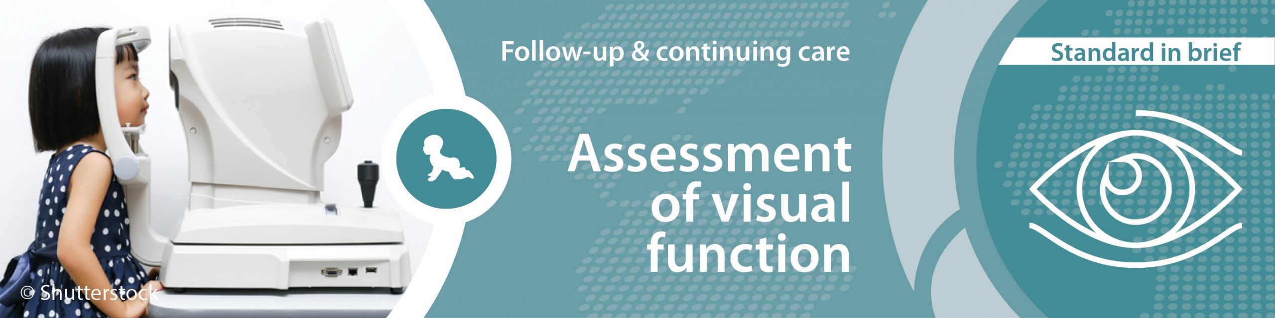
Target group
Infants born very preterm or those infants with risk factors (see preamble Follow-up & continuing care) and parents
User group
Healthcare professionals, neonatal units, hospitals, follow-up teams, and health services
Statement of standard
Standardised visual assessment is conducted by age 3.5 to 4 years and repeated by age 5 to 6 (1), at which age additional attention is payed to visual information processing dysfunctions.
Rationale
The goal is to assess and evaluate the development of visual and visual information processing functions to identify those who could benefit from additional support and early intervention.
Preterm born infants have an increased risk of visual dysfunctions, in particular those with severe brain injury and those who suffered from severe and/or treated retinopathy of prematurity (ROP). Long-term follow-up showed that an adverse ophthalmological outcome (AOO) (reduced acuity, strabismus, high myopia, colour defect, field defect and/or subnormal contrast sensitivity) is present in 25-50% of preterm infants with a birth weight <1500 g. (2,3)Infants who suffered from grade 2 or 3 hypoxic ischaemic encephalopathy or meningoencephalitis have an increased risk of (cerebral) visual impairment (CVI) (7-11% and 17% respectively). (4–6)Impairments include dysfunctions in visual sensory, oculomotor and perceptive (such as object recognition and spatial processing) functioning. Both visual sensory and visual perceptive dysfunctions exert a negative effect on visuomotor, neuropsychological outcome and academic skills such as reading, writing and maths achievement and on quality of life. (7–11)
Benefits
Short-term benefits
In infants with co-morbidities, early assessment and diagnosis could lead to early visual intervention, proactive treatment, and support.
Long-term benefits
Early diagnosis of visual impairment promotes timely interventions (12,13)
Promotes realistic expectations in those with severe impairment (consensus)
Improved decision making for schooling and learning support (consensus)
Provides feedback to perinatal and neonatal services and healthcare officials (consensus)
Reduced risk of misdiagnoses (e.g. reading difficulties) (consensus)
Improved parental awareness and parent-infant interaction adapted to visual ability (consensus)
Improved academic outcome (14)
Improved social integration and quality of life (3,11)
Reduced social burden and costs (consensus)
Components of the standard
Component
Grading of evidence
Indicator of meeting the standard
For parents and family
Parents are informed about and invited, in a timely manner, by healthcare professionals to attend follow-up programme, which includes visual assessments (define ages at which visual follow-up should take place and the provider thereof). (3)
A (High quality) B (High quality)
Patient information sheet1
Parents receive standardised feedback about the results of their child’s visual health screening in a language that is accessible to them.
B (High quality)
Parent feedback
Parents are informed about the need for early intervention and treatment and support options in case of visual impairments.
B (High quality)
Patient information sheet1
Parents are asked for permission to allow their child’s medical and educational information to be used by healthcare professionals for outcome measures.
B (Low quality)
Parent consent, patient information sheet1
Parents are asked to consent to share the results of their child’s visual screening tests with education services.
B (Moderate quality)
Parent consent
For healthcare professionals
A guideline on follow-up programme including visual assessment is adhered to by all healthcare professionals.
B (High quality)
Guideline
Regular training on standardised visual assessment in high-risk infants in which gestational age, ROP status, and brain damage are taken into account is attended by all responsible healthcare professionals. (2,3,15–17)
A (High quality) B (High quality)
Training documentation
Children with ROP grade ≤2 undergo ophthalmologic screening at 3.5-4 years and assessment of visual acuity at 4-5 years; at younger ages, children with signs of adverse visual development are referred directly to the paediatric ophthalmologist. (2,3,14,17)
A (High quality) B (High quality)
Guideline
Children with ROP grades 3 and 4 (or treated for any grade of ROP) and/or with severe brain damage have regular follow-up assessments at the discretion of the ophthalmologist and are at least screened for strabismus and refractive errors at 12 months. (18)
A (High quality)
Guideline
Children with clinical suspicion for visual perception dysfunctions are assessed at 3 years of age onwards, based on psychometrically sound assessment tools. (11,19)
A (High quality)
Audit report2, guideline
For neonatal unit, hospital and follow-up team
A guideline on follow-up programme including visual assessment is available and regularly updated.
B (High quality)
Guideline
A follow-up programme after discharge including visual assessment, and referral and signposting to supportis funded, provided,and supported.
B (Moderate quality)
Audit report2
Training on standardised visual assessment in high-risk infants is ensured.
B (High quality)
Training documentation
For health service
A national guideline on follow-up programme including visual assessment is available and regularly updated.
B (High quality)
Guideline
A follow-up service including visual assessment and intervention is specified, provided, funded and monitored.
B (Moderate quality)
Audit report2
1The indicator “patient information sheet” is an example for written, detailed information, in which digital solutions are included, such as web-based systems, apps, brochures, information leaflets, and booklets.
2The indicator “audit report” can also be defined as a benchmarking report.
Where to go
Further development
Grading of evidence
For parents and family
Offer visual follow-up until adult age. (20)
B (Moderate quality)
Families are supported by case manager in order to ensure follow-up programme, which includes visual assessments, and signposting to regular support is provided.
B (High quality)
For healthcare professionals
N/A
For neonatal unit and follow-up team
Establish an integrated electronic system with follow-up provider to schedule follow-up visits.
B (Moderate quality)
For hospital and follow-up team
Establish multidisciplinary teams, including ophthalmologist/neuropsychologist specialised in visual perception, to evaluate high-risk children. (3)
B (Moderate quality)
For health service
Support the development of reliable and valid instruments to assess cerebral visual deficits with country specific norms and facilitate differential diagnosis. (15,19)
A (High quality) B (High quality)
Develop a national network for benchmarking of follow-up quality and provision of care.
B (Moderate quality)
Getting started
Initial steps
For parents and family
Parents are informed as soon as possible, by healthcare professionals about the risks to vision after high-risk birth and about the follow-up programme.
For healthcare professionals
Attend appropriate and regular training on standardised visual assessment.
Establish a structure of communication with other healthcare institutions, who are providing follow-up care.
For neonatal unit and follow-up team
Develop and implement a guideline on follow-up programmes, which include visual assessment.
Develop information material for parents to raise awareness about the importance of visual follow-up assessment.
Establish at least a standardised formal system of keeping track of families.
Develop a structure for local follow-up care.
For hospital and follow-up team
Support healthcare professionals to participate in regular training on standardised visual assessments.
Ensure ophthalmologists are available and regularly trained in visual sequelae of high-risk births.
For health service
Develop and implement a national guideline on follow-up programmes which include visual assessment.
Make a policy decision that visual follow-up services is standard of care for all infants.
Retinopathy of prematurity (ROP) is an important cause of visual impairment in the preterm infant, and is due to disorganized vascular development of the retina usually after retinal ischaemia consequent to oxygen exposure. Infants who develop ROP are at increased risk of ophthalmological deficits such as refractive error (up to 64%), amblyopia and strabismus (36-44%). (21)However, these disorders are also prevalent in those born under 32 weeks without ROP, in whom refractive errors are present in 26% of infants, amblyopia in 21% and strabismus in 16-20%. (15) In preterm children attending mainstream school, decreased visual acuity was reported to occur two to three times more frequently than in term-born peers, principally due to refractive errors. High myopia and anisometropia, in particular, confer a risk for developing amblyopia and strabismus. Such early reductions of visual acuity are reportedly subject to “catch-up” by age 3.5-4 years, following timely treatment. (21) Weight at birth, head circumference at birth and head circumference at 5,5 years seem to be important contributing factors. (22)
Premature infants are born in a phase of rapid brain growth and organisation. Alterations of brain development have been shown in the neonatal period but can last into adulthood, both in structure, altered networks and function, also in the visual areas of the brain. (23–28) Visual impairments caused by adverse brain development are collectively referred to as cerebral visual impairment (CVI) and include both visual sensory impairment and deficient visual perception. CVI nowadays is the most frequent cause of visual impairment in children in developed countries, in contrast to the visual sequelae of ROP (29) , and is associated with deficiencies in the development of cognition and motor abilities. (15,30,31) CVI covers a wide range of deficits, from children merely suffering from spatial processing dysfunctions to deficits in object recognition and scene identification, and also cortically blind children, having no visual perception at all. (11,15)
In preterm born children, CVI is typically diagnosed in children with periventricular white matter disease, thus particularly in those born <32 weeks of gestation, although its prevalence is not exactly known. (32) However, CVI can also emerge in children without evident/overt brain pathology. The clinical profile of visual perceptive deficits can change during childhood. (15)Once CVI is suspected, regular follow-up of visual functioning is therefore advised.
Severe visual sensory and oculomotor deficits mostly become visible at early ages. However, visual screening is most reliable at the age of 3.5 to 4 years. At 5 to 6 years, most visual sensory and oculomotor problems have become apparent. In case of suspicion of visual perceptive dysfunctions, screening is feasible in infants.(33) Neuropsychological batteries for diagnosing CVI from the age of 3 years onward have been developed. (34) Furthermore, (inter)national guidelines for diagnosis and referral in case of suspicion of CVI, have recently been published. (35,36) At least those children affected by brain damage, should undergo the described work-up for CVI. Refractive error can often be corrected. Strabismic amblyopia needs to be corrected at an early stage with patching. The treatment or support of visual perceptual deficits aims to offer the child the best environment to improve its visual functioning and to learn strategies to cope with its specific deficits. Micheletti et al nicely summarised the evidence for early identification of visual dysfunctions, enabling access to early visual intervention, which may support visual and overall development. (13)
Horwood A, Heijnsdijk E, Kik J, Sloot F, Carlton J, Griffiths HJ, et al. A population-level post-screening treatment cost framework to help inform vision screening choices for children under the age of seven. Strabismus. 2023 Sep;31(3):220–35.
Stephenson T, Wright S, O’Connor A, Fielder A, Johnson A, Ratib S, et al. Children born weighing less than 1701 g: visual and cognitive outcomes at 11-14 years. Arch Dis Child Fetal Neonatal Ed. 2007 Jul;92(4):F265-270.
Holmström G, Larsson E. Long-term follow-up of visual functions in prematurely born children–a prospective population-based study up to 10 years of age. J AAPOS Off Publ Am Assoc Pediatr Ophthalmol Strabismus. 2008 Apr;12(2):157–62.
Stevens JP, Eames M, Kent A, Halket S, Holt D, Harvey D. Long term outcome of neonatal meningitis. Arch Dis Child Fetal Neonatal Ed. 2003 May;88(3):F179-184.
Azzopardi D, Strohm B, Marlow N, Brocklehurst P, Deierl A, Eddama O, et al. Effects of hypothermia for perinatal asphyxia on childhood outcomes. N Engl J Med. 2014 Jul 10;371(2):140–9.
Williams C, Warnes P, Jary S, Young G, Blair PS, Benton CP, et al. Vision function in children 10 years after grade 3 or 4 intraventricular haemorrhage with ventricular dilation: A masked prospective study. Dev Med Child Neurol. 2023 Feb;65(2):223–31.
Molloy CS, Di Battista AM, Anderson VA, Burnett A, Lee KJ, Roberts G, et al. The contribution of visual processing to academic achievement in adolescents born extremely preterm or extremely low birth weight. Child Neuropsychol J Norm Abnorm Dev Child Adolesc. 2017 Apr;23(3):361–79.
Rapin I. Dyscalculia and the Calculating Brain. Pediatr Neurol. 2016;61:11–20.
Downie ALS, Jakobson LS, Frisk V, Ushycky I. Periventricular brain injury, visual motion processing, and reading and spelling abilities in children who were extremely low birthweight. J Int Neuropsychol Soc JINS. 2003 Mar;9(3):440–9.
Beligere N, Perumalswamy V, Tandon M, Mittal A, Floora J, Vijayakumar B, et al. Retinopathy of prematurity and neurodevelopmental disabilities in premature infants. Semin Fetal Neonatal Med. 2015 Oct;20(5):346–53.
Ortibus E, Fazzi E, Dale N. Cerebral Visual Impairment and Clinical Assessment: The European Perspective. Semin Pediatr Neurol. 2019 Oct;31:15–24.
Chavda S, Hodge W, Si F, Diab K. Low-vision rehabilitation methods in children: a systematic review. Can J Ophthalmol J Can Ophtalmol. 2014 Jun;49(3):e71-73.
Micheletti S, Merabet LB, Galli J, Fazzi E. Visual intervention in early onset visual impairment: A review. Eur J Neurosci. 2023 Jun;57(12):1998–2016.
Holmström G, Larsson E. Outcome of retinopathy of prematurity. Clin Perinatol. 2013 Jun;40(2):311–21.
Ortibus EL, De Cock PP, Lagae LG. Visual perception in preterm children: what are we currently measuring? Pediatr Neurol. 2011 Jul;45(1):1–10.
Ricci D, Romeo DM, Gallini F, Groppo M, Cesarini L, Pisoni S, et al. Early visual assessment in preterm infants with and without brain lesions: correlation with visual and neurodevelopmental outcome at 12 months. Early Hum Dev. 2011 Mar;87(3):177–82.
Hellström A, Källén K, Carlsson B, Holmström G, Jakobsson P, Lundgren P, et al. Extreme prematurity, treated retinopathy, bronchopulmonary dysplasia and cerebral palsy are significant risk factors for ophthalmological abnormalities at 6.5 years of age. Acta Paediatr Oslo Nor 1992. 2018 May;107(5):811–21.
AMERICAN ACADEMY OF PEDIATRICS Section on Ophthalmology, AMERICAN ACADEMY OF OPHTHALMOLOGY, AMERICAN ASSOCIATION FOR PEDIATRIC OPHTHALMOLOGY AND STRABISMUS, AMERICAN ASSOCIATION OF CERTIFIED ORTHOPTISTS. Screening Examination of Premature Infants for Retinopathy of Prematurity. PEDIATRICS. 2013 Jan 1;131(1):189–95.
Geldof CJA, van Wassenaer-Leemhuis AG, Dik M, Kok JH, Oosterlaan J. A functional approach to cerebral visual impairments in very preterm/very-low-birth-weight children. Pediatr Res. 2015 Aug;78(2):190–7.
Darlow BA, Elder MJ, Kimber B, Martin J, Horwood LJ. Vision in former very low birthweight young adults with and without retinopathy of prematurity compared with term born controls: the NZ 1986 VLBW follow-up study. Br J Ophthalmol. 2017 Dec 6.
Fierson WM, American Academy of Pediatrics Section on Ophthalmology, American Academy of Ophthalmology, American Association for Pediatric Ophthalmology and Strabismus, American Association of Certified Orthoptists. Screening examination of premature infants for retinopathy of prematurity. Pediatrics. 2013 Jan;131(1):189–95.
Raffa L, Aring E, Dahlgren J, Karlsson AK, Andersson Grönlund M. Ophthalmological findings in relation to auxological data in moderate-to-late preterm preschool children. Acta Ophthalmol (Copenh). 2015 Nov;93(7):635–41.
Brumbaugh JE, Conrad AL, Lee JK, DeVolder IJ, Zimmerman MB, Magnotta VA, et al. Altered brain function, structure, and developmental trajectory in children born late preterm. Pediatr Res. 2016;80(2):197–203.
Groppo M, Ricci D, Bassi L, Merchant N, Doria V, Arichi T, et al. Development of the optic radiations and visual function after premature birth. Cortex. 2014 Jul 1;56:30–7.
Kelly CE, Cheong JLY, Molloy C, Anderson PJ, Lee KJ, Burnett AC, et al. Neural Correlates of Impaired Vision in Adolescents Born Extremely Preterm and/or Extremely Low Birthweight. PLOS ONE. 2014 Mar 24;9(3):e93188.
Pavaine J, Young JM, Morgan BR, Shroff M, Raybaud C, Taylor MJ. Diffusion tensor imaging-based assessment of white matter tracts and visual-motor outcomes in very preterm neonates. Neuroradiology. 2016 Mar;58(3):301–10.
Ramenghi LA, Ricci D, Mercuri E, Groppo M, De Carli A, Ometto A, et al. Visual performance and brain structures in the developing brain of pre-term infants. Early Hum Dev. 2010 Jul;86 Suppl 1:73–5.
Thompson DK, Thai D, Kelly CE, Leemans A, Tournier JD, Kean MJ, et al. Alterations in the optic radiations of very preterm children-Perinatal predictors and relationships with visual outcomes. NeuroImage Clin. 2014;4:145–53.
Bunce C, Xing W, Wormald R. Causes of blind and partial sight certifications in England and Wales: April 2007-March 2008. Eye Lond Engl. 2010 Nov;24(11):1692–9.
Geldof CJA, van Hus JWP, Jeukens-Visser M, Nollet F, Kok JH, Oosterlaan J, et al. Deficits in vision and visual attention associated with motor performance of very preterm/very low birth weight children. Res Dev Disabil. 2016 Jul;53–54:258–66.
Geldof CJA, van Wassenaer AG, de Kieviet JF, Kok JH, Oosterlaan J. Visual perception and visual-motor integration in very preterm and/or very low birth weight children: a meta-analysis. Res Dev Disabil. 2012 Apr;33(2):726–36.
Dutton GN, McKillop ECA, Saidkasimova S. Visual problems as a result of brain damage in children. Br J Ophthalmol. 2006 Aug;90(8):932–3.
Rossi A, Gnesi M, Montomoli C, Chirico G, Malerba L, Merabet LB, et al. Neonatal Assessment Visual European Grid (NAVEG): Unveiling neurological risk. Infant Behav Dev. 2017 Nov;49:21–30.
Vancleef K, Janssens E, Petré Y, Wagemans J, Ortibus E. Assessment tool for visual perception deficits in cerebral visual impairment: reliability and validity. Dev Med Child Neurol. 2020 Jan;62(1):118–24.
Boonstra FN, Bosch DGM, Geldof CJA, Stellingwerf C, Porro G. The Multidisciplinary Guidelines for Diagnosis and Referral in Cerebral Visual Impairment. Front Hum Neurosci. 2022;16:727565.
Pilling RF, Allen L, Bowman R, Ravenscroft J, Saunders KJ, Williams C. Clinical assessment, investigation, diagnosis and initial management of cerebral visual impairment: a consensus practice guide. Eye Lond Engl. 2023 Jul;37(10):1958–65.
Second edition, December 2024. Previous edition reviewed by Professor Nikolaos Kozeis.
Lifecycle
5 years/next revision: 2029
Recommended citation
GFCNI, Ortibus E, Huening B et al., European Standards of Care for Newborn Health: Assessment of visual function. 2024.
Please enter your job description and origin country
You are currently viewing a placeholder content from Facebook. To access the actual content, click the button below. Please note that doing so will share data with third-party providers.
You are currently viewing a placeholder content from Instagram. To access the actual content, click the button below. Please note that doing so will share data with third-party providers.
You are currently viewing a placeholder content from X. To access the actual content, click the button below. Please note that doing so will share data with third-party providers.